Google Maps

 Waze Maps
 Apple Maps
 All calls are free of charge
 765 22 88
 765 22 88
 765 22 88
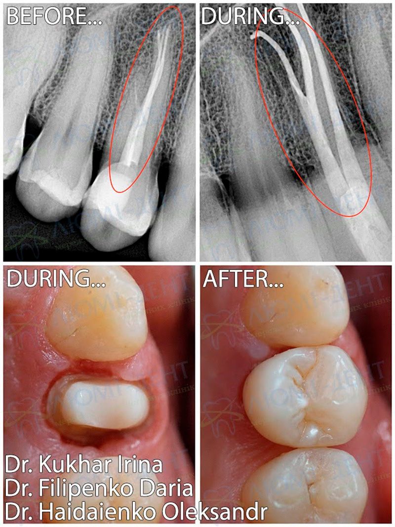


In this clinical case, the patient presented with persistent aching pain in the area of tooth 1.4 that had continued for two weeks after treatment performed at another dental clinic.
During the examination and CBCT scan of the jaws, apical periodontitis of tooth 1.4 was diagnosed. The cause of the condition was a previously missed root canal.
Before treatment, professional oral hygiene, a CBCT scan, and periapical radiography were performed. Treatment was carried out under STA computer-assisted anesthesia with rubber dam isolation.
After removal of the filling and retreatment of the root canals, a previously missed canal was identified. Under the control of a Seiler Evolution XR6 operating microscope, mechanical and chemical preparation of the root canal system was performed using NiC and SOCO instruments with ultrasonic-activated irrigation.
Following hermetic obturation of the root canals using the warm gutta-percha 3D technique, the tooth was restored with Estelite Asteria composite material and prepared for the prosthetic stage of treatment.
The final stage involved fabrication and placement of a full-anatomical zirconia crown using CAD/CAM technology, which allowed complete restoration of the tooth’s function and aesthetics.
Result:
The source of infection and pain was eliminated, while full tooth function and a long-term aesthetic result were restored.
Discover the advanced dental care available at our clinic.
Who provided the treatment:
|
The endodontic stage was performed by dentist-therapist, endodontist
|
Periodontal support was provided by periodontist
|
The orthopedic stage was performed by prosthodontist
|