Google Maps

 Waze Maps
 Apple Maps
 All calls are free of charge
 765 22 88
 765 22 88
 765 22 88
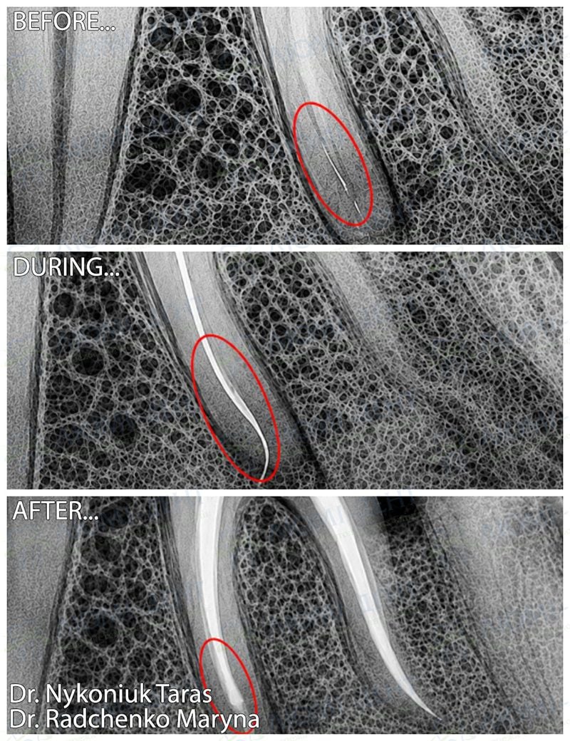


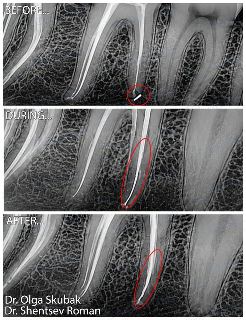
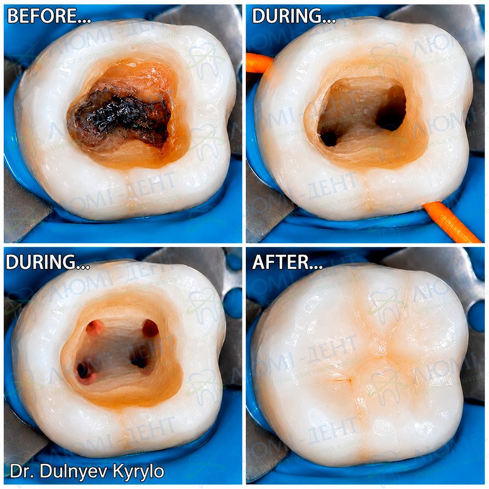
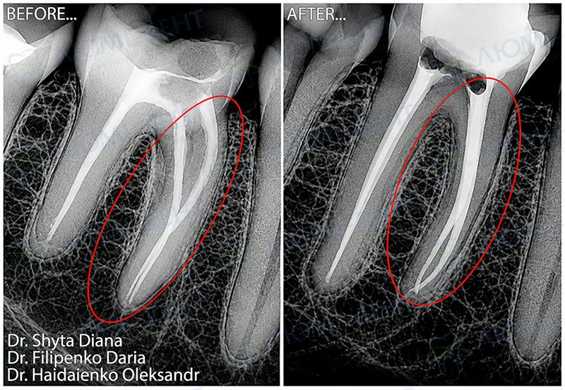
In this case, the patient was referred for root canal retreatment after unsuccessful treatment at another clinic. An X-ray revealed a fractured instrument fragment in one of the root canals.
The examination confirmed apical periodontitis of tooth 3.6.
For treatment planning, a CBCT scan was performed to determine the exact position of the fragment and assess the root canal anatomy.
Using a Leica M320 dental microscope, the fractured instrument was bypassed with the ByPass technique, followed by mechanical and chemical canal preparation and obturation with warm gutta-percha.
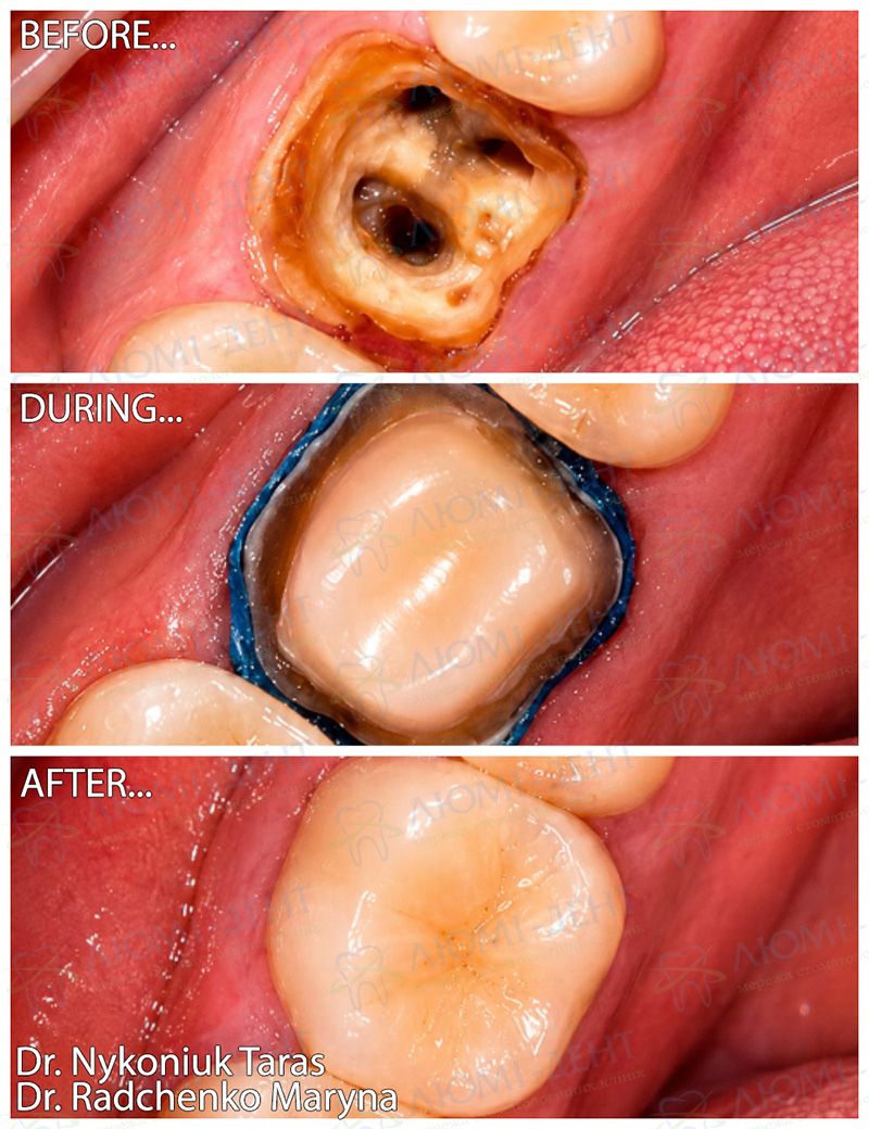
After endodontic treatment, the tooth was restored with composite material and a zirconia crown fabricated using CAD/CAM technology.
Result:
The inflammatory process has been eliminated, the tooth preserved, and chewing function restored.
Learn more about our clinic and advanced dental treatment options by following this link.
Who performed the treatment:
|
The endodontic stage was performed by dentist-therapist
|
The prosthetic stage was performed by prosthodontist
|