Google Maps

 Waze Maps
 Apple Maps
 All calls are free of charge
 765 22 88
 765 22 88
 765 22 88
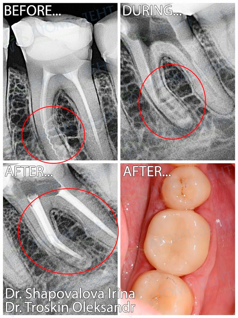


In this clinical case, the patient presented with periodic pain in tooth 3.6.
During the examination and CBCT scan of the jaws, apical periodontitis of tooth 3.6 was diagnosed.
Treatment was performed under STA computer-assisted anesthesia with rubber dam isolation. Using a Leica M320 operating microscope, the root canals were retreated, a fractured endodontic instrument fragment was removed, and mechanical and chemical preparation of the canals was performed with ultrasonic-activated irrigation.
After hermetic root canal filling using the warm gutta-percha 3D obturation technique, the tooth was restored with Estelite Asteria composite material and prepared for the prosthetic stage.
The final stage included digital scanning, fabrication, and placement of a full-anatomical zirconia crown using CAD/CAM technology.
Result:
Pain was eliminated, and the tooth was fully restored and reliably protected.
Learn why patients trust our clinic for advanced dental treatment.
Who provided the treatment:
|
The endodontic stage was performed by dentist-therapist, endodontist
|
The orthopedic stage was performed by prosthodontist
|